
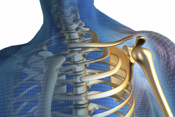
What is calcific tendonitis?
The degenerative changes, which occur in tendons as part of the aging process can, in combination with exertion, cause chronic inflammation with deposits of calcium in the supraspinatus tendon. These calcium deposits can rupture into the bursa overlying the supraspinatus tendon, bringing about a temporary improvement of the condition but causing Bursitis.
Symptoms can be intense pain suddenly on the front of the shoulder, which can be so severe it prevents sleep.
Causes
No one is sure why some people develop calcific tendonitis but it is more common in people who play sports or routinely raise their arms up and down for work but calcific tendonitis can affect anyone.
Most commonly seen in adults between 40-60 years old with women much more likely to get it then men.
Other causes can be:
-A genetic predisposition
-Abnormal cell growth
-Abnormal thyroid gland activity
-bodily production of anti-inflammatory agents
-metabolic diseases, such as diabetes
An Xray can reveal larger deposits, and ultrasound is also a useful tool to locate smaller deposits.
Most cases of calcific tendonitis can be treated without surgery. In mild cases, your doctor may recommend a mix of medication and physiotherapy or a nonsurgical procedure.
Medication
Non-steroidal anti-inflammatory drugs (NSAIDs) are considered to be the first line of treatment. These medications are available over the counter and include:
- aspirin
- ibuprofen
- naproxin
Be sure to follow the recommended dosing on the label, unless your doctor advises otherwise.
Your doctor may also recommend a cortisone injection to help relieve any pain or swelling.
Nonsurgical procedures
In mild-to-moderate cases, your doctor may recommend one of the following procedures.
Extracorporeal shock-wave therapy (ESWT): The technician will use a small handheld device to deliver mechanical shocks to your shoulder, near the site of calcification.
Higher frequency shocks are more effective but can be painful. They can adjust the shock waves to a level you can tolerate.
This therapy may be performed once a week for 3 weeks.
Radial shock-wave therapy (RSWT):
The technician will use a handheld device to deliver low- to medium-energy mechanical shocks to the affected part of the shoulder. This produces effects similar to ESWT.
Therapeutic ultrasound: The physiotherapist will use a handheld device to direct a high frequency sound wave at the calcific deposit. This helps break down the calcium crystals and is usually painless.
Percutaneous needling: This therapy is more invasive than other nonsurgical methods. After administering local anaesthesic to the area, your doctor will use a needle to make small holes in your skin. This will allow them to manually remove the deposit. This may be done in conjunction with ultrasound to help guide the needle into the correct position.
Surgery
About 10% of people will need surgery to remove the calcium deposit.
This is normally done arthroscopically via a small incision and a tiny camera. The camera will guide the surgical tool to help locate and guide the tool to remove the deposit.
Your recovery period will depend on the size, location, and number of calcium deposits. Some people will return to normal functioning within a week and others may experience post-surgical pain which continues to limit their activities.
You will be sent to physiotherapy after surgery.
Physiotherapy
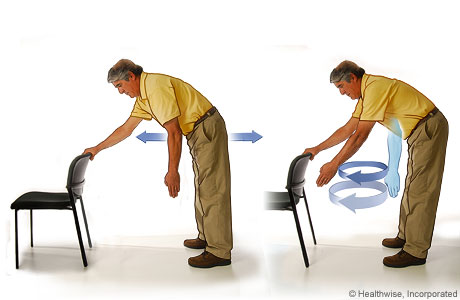
You will be shown the correct exercises by the physiotherapist to regain the full range of motion of your shoulder.
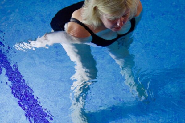
These may include some easy pendulum swinging exercises that do not cause any pain and ease the stiffness in the shoulder. Hydrotherapy can be another easy way to exercise and regain movement.
see Hydrotherapy
Rehabilitation after surgery
Recovery time after surgery varies from person to person and may take up to 3 months
Recovery from arthroscopic surgery is generally quicker than from open surgery.
After either open or arthroscopic surgery, your doctor may advise you to wear a sling for a few days to support and protect the shoulder.
You should also expect to attend physiotherapy sessions for 6-8 weeks. Physiotherapy usually begins with some stretching and very limited range-of-motion exercises. You will typically progress to some light weight-bearing activity after four weeks.
Prognosis
Although calcific tendonitis can painful for some, a quick resolution is likely and only 10% of people require some form of surgery.
Calcific tendonitis does eventually disappear on its own but can lead to complications if left untreated.